825 18th Street
Vero Beach, FL 32960

How We Help Pets
A diagnosis is only the beginning of the story.
This resource is designed to give pet owners information, direction, and support. We are only one part of your pet's journey, and our role is to work alongside your primary veterinarian, specialists, and family to help your pet move, feel, and function as well as possible.
How We Help Pets
Pet owners often arrive with a diagnosis, but a diagnosis alone does not always tell you what daily life will look like, what changes to watch for, what you can do at home, or how rehabilitation may fit into the larger care plan.
Our goal is to provide practical information, direction, and support. We look at the whole pet: comfort, mobility, strength, neurologic function, weight, home environment, medications and supplements, family goals, and the activities that matter most to that individual pet.
Success is not defined by perfection. We want pets to move as comfortably, confidently, and efficiently as possible while minimizing unnecessary compensation.

Dr. Amber's Tip
A diagnosis is only the beginning of the story. We are only part of the journey.
Medical References & Resources
Healthy Aging, Pain & Wellness
These pages focus on proactive care, mobility, strength, body condition, cognition, and helping pets continue to enjoy more good days as they age.
Healthy aging is not a one-time plan. Bodies change, needs change, and plans should be periodically rebalanced.
Medical References & Resources
Arthritis
Arthritis is a chronic, progressive joint disease that causes pain, stiffness, reduced range of motion, and changes in how a pet moves.
Learn More
Healthy Aging
Healthy aging is proactive care for the whole pet. The goal is not simply to help pets live longer, but to help them live better.
We monitor mobility, muscle mass, balance, pain, body condition, cognition, endurance, home needs, medications, supplements, and quality of life. The plan is periodically rebalanced because an aging body does not stay the same.
What might you notice at home?
Slower recovery after activity
Subtle changes in stairs, jumping, or rising
Loss of muscle
Weight change
Changes in sleep or behavior
Reduced confidence on slippery floors
What Do We Do?
Mobility and pain assessment
Strength and conditioning
Body-condition and nutrition support
Medication and supplement review
Home modifications
Brain-health and enrichment planning
Periodic rebalancing as needs change
Dr. Amber's Tip
The best time to build a healthy-aging plan is before a crisis. Small changes are easier to address when we notice them early.
Hope for Your Pet
Healthy aging is about preserving comfort, independence, engagement, and the activities that make your pet's life meaningful.
Medical References & Resources
Sarcopenia
Sarcopenia is the loss of skeletal muscle mass and strength that commonly occurs with aging, illness, inactivity, and chronic pain.
Muscle is not just cosmetic. It supports joints, balance, endurance, metabolism, and the ability to rise, walk, climb, and recover from illness or surgery.
What might you notice at home?
Thinner thighs or shoulders
Prominent spine or hips
Difficulty rising
Reduced endurance
Slower walking
Loss of balance or confidence
What Do We Do?
Progressive strengthening
Safe conditioning
Protein and nutrition discussion with the veterinary care team
Weight and body-condition monitoring
Balancing supplements and medications when appropriate
Home exercise that is sustainable for the pet and family
Dr. Amber's Tip
Watch muscle, not just body weight. A pet can maintain the same number on the scale while losing important lean muscle.
Hope for Your Pet
Preserving muscle is one of the most useful ways we can support mobility and independence as pets age.
Medical References & Resources
Weight Loss & Body Condition
Maintaining a lean body condition is one of the most effective ways to reduce stress on the body, slow progression of many mobility problems, and support a longer, more comfortable life.
Successful weight loss is not about finding a perfect diet. It is about finding the right fit for your dog or cat: a diet that supports their medical and nutritional needs, is sustainable for the family, and agrees with the pet.
Diet is the primary driver of weight loss. Exercise is a secondary component, but it is vital for building a healthy, conditioned, comfortable body.
What might you notice at home?
Loss of a visible waist
Difficulty feeling the ribs
Reduced endurance
Difficulty rising or climbing
Exercise intolerance
Worsening joint or back discomfort
What Do We Do?
Realistic target weight and body-condition goals
Nutrition planning with the veterinary team
Safe exercise based on current mobility
Strength and conditioning while weight is changing
Regular weigh-ins and plan adjustments
Dr. Amber's Tip
The best weight-loss plan is the one your pet and family can actually maintain.
Hope for Your Pet
Weight management is not about appearance. It is about reducing unnecessary load on joints and helping the body function as efficiently as possible.
Medical References & Resources
Cognitive Dysfunction Syndrome (CDS)
Cognitive dysfunction syndrome (CDS) is a common age-related disease that affects the brain, causing deterioration that is often compared with Alzheimer's disease in people.
Changes can be reported in cats around 10 years of age and in dogs as young as 9 years. Because early signs may be subtle, families often assume the changes are simply part of getting older.
What might you notice at home?
Disorientation
Changes in sleep-wake cycles
House-soiling changes
Altered interaction with family
Anxiety or restlessness
Changes in learned behaviors or activity
What Do We Do?
Cognitive screening and monitoring
Environmental enrichment
Predictable routines
Safe physical activity
Home modifications
Discussion of nutrition, supplements, and medications with the veterinary care team
Periodic rebalancing as signs change
Dr. Amber's Tip
Behavior change in an older pet deserves a medical conversation. Do not assume every change is simply aging.
Hope for Your Pet
Supporting the brain, body, environment, and family together can help older pets remain engaged and comfortable.
Medical References & Resources
Orthopedic Conditions & Surgical Recovery
Orthopedic rehabilitation focuses on comfort, mechanics, strength, range of motion, confidence, and reducing compensatory movement patterns.
Medical References & Resources
Gracilis Injury & Fibrotic Myopathy
The gracilis is a large muscle along the inside of the thigh that helps bring the rear leg toward the body and contributes to movement of the hip and stifle.
Like any muscle, the gracilis can be injured. Acute injury can cause pain, inflammation, and scar formation during healing. Gracilis fibrotic myopathy is a distinct chronic condition in which muscle is progressively replaced by dense, less-flexible fibrous tissue. As the muscle becomes restricted, the rear-limb gait can become very characteristic.
German Shepherd Dogs, particularly working dogs, are overrepresented. Repetitive muscular stress or microtrauma and genetic predisposition have been proposed, but the exact cause of fibrotic myopathy is not completely understood.
What might you notice at home?
Rear-leg pain or lameness after an acute injury
Tenderness or changes in the inner thigh
Shortened stride
Recurring lameness with activity
With established fibrosis: an abrupt inward movement of the limb or paw during swing
What Do We Do?
With earlier muscle injury: manage comfort, maintain appropriate flexibility, use controlled loading, support scar remodeling, and develop a gradual return to activity.
With established fibrotic myopathy: maintain comfortable mobility, strengthen surrounding muscles, improve movement efficiency, reduce unnecessary compensation, and preserve conditioning.
Rehabilitation cannot turn mature fibrous tissue back into normal muscle, so goals change as the condition becomes chronic.
Dr. Amber's Tip
Timing changes our goals. Early on we may be able to influence healing and preserve flexibility. With established fibrosis, we focus on function, conditioning, and how the rest of the body moves around the restriction.
Hope for Your Pet
A gracilis injury does not automatically become fibrotic myopathy. When chronic fibrosis is present, rehabilitation can still support efficient movement, conditioning, comfort, and long-term function.
Medical References & Resources
Common Calcaneal (Achilles) Tendon Injury
An injury to the Achilles - more accurately called the common calcaneal tendon - can be startling to see and may be painful. The common calcaneal tendon is a group of tendons working together at the back of the hock (ankle) to support normal standing and movement.
In dogs, injury may occur suddenly from trauma or develop more gradually from chronic degeneration. The tendon can be partially injured or completely disrupted. The position of the hock and toes can give clues about which tendon components remain intact.
What might you notice at home?
Swelling or pain around the tendon
Rear-leg lameness
The hock dropping closer to the ground
A flat or plantigrade stance
Toes curling downward
Shortened or abnormal stride
Difficulty pushing off the affected leg
What Do We Do?
Protect the healing tendon and respect surgical or external-support restrictions.
Maintain appropriate mobility in surrounding joints.
Reduce muscle loss during restricted activity.
Gradually rebuild strength, stability, body awareness, and weight bearing.
Address compensatory movement.
Develop a staged return-to-activity plan.
Dr. Amber's Tip
Tendon healing and feeling better don't happen at the same speed. A pet may want to run long before the healing tendon is ready for that load.
Hope for Your Pet
Whether treatment is surgical or nonsurgical, rehabilitation supports the whole limb through pain, muscle loss, compensation, stability, confidence, and eventual return to activity.
Medical References & Resources
Fractured Digits & Toe Tumors
A painful toe may seem like a small problem, but dogs and cats place significant force through their feet with every step. Digits can fracture during running, jumping, rough play, falls, or other trauma.
Not every swollen or painful toe is an injury. Persistent swelling, pain, nail changes, or lameness can also be caused by infection, inflammatory or immune-mediated disease, or a tumor involving the digit or nail bed.
Changes to the nail or nail bed - including abnormal nail growth, repeated breakage, bleeding, swelling, or unexplained loss of a toenail - deserve evaluation.
What might you notice at home?
Sudden or persistent limping
Holding up a paw
Swelling of one toe
Pain when the foot is touched
Licking or chewing the foot
Broken, abnormal, or repeatedly lost nail
Bleeding or drainage around the nail
A toe that changes size or shape
A problem that improves but repeatedly returns
What Do We Do?
For fractures: support comfortable weight bearing, protect healing tissues, maintain strength, and gradually return to activity.
After digit amputation: help the pet adapt to altered foot mechanics, rebuild confidence, and reduce compensatory loading.
Monitor how the remaining toes and pads accept weight.
Maintain strength in the affected limb and protect other limbs from unnecessary compensation.
Use appropriate flooring, exercise progression, and foot care.
Dr. Amber's Tip
Pay attention to the toenails. A toenail that falls off, repeatedly breaks, grows abnormally, bleeds, or has persistent swelling around the nail bed may be telling us something about the health of the toe - or sometimes the health of the pet as a whole. If you don't know why a toenail was lost, it's worth asking why.
Hope for Your Pet
A fractured toe can heal, and even when a tumor requires digit amputation, many dogs and cats adapt very well. Our goal is to help them move comfortably and confidently rather than simply leaving them to figure out the new mechanics on their own.
Medical References & Resources
Limber Tail - When the Wag Suddenly Stops
Dogs use their tails for communication, balance, movement, and interaction with the world. Sometimes after vigorous activity, swimming, hunting, rough play, cold-weather exposure, or prolonged confinement, the muscles controlling the tail can become acutely injured and painful.
This condition is commonly called limber tail, swimmer's tail, or cold tail. The medical term is acute caudal myopathy.
Not every limp tail is limber tail. Tail fracture or dislocation, anal gland disease, lower-back or spinal disease, nerve injury, infection, and other problems can also change tail position, so a first episode deserves veterinary evaluation.
What might you notice at home?
Tail hanging limp from the base
Tail extending normally for a short distance and then dropping
Reduced wagging
Pain near the tail base
Difficulty sitting comfortably
Reluctance to squat
What Do We Do?
Appropriate activity modification and rest.
Gentle soft-tissue care when appropriate and comfortable.
Address secondary muscle tension.
Maintain comfortable movement.
Gradually return to regular activity as signs resolve.
Dr. Amber's Tip
A tail that suddenly stops wagging is worth paying attention to. First make sure the problem really is the tail muscles - and not the bones, nerves, anal glands, or spine.
Hope for Yur Pet
Uncomplicated limber tail is usually temporary, and many dogs regain their usual tail movement over several days. Sometimes the missing wag looks much scarier than the injury ultimately turns out to be.
Medical References & Resources
Spondylosis Deformans
Spondylosis deformans is often described as 'arthritis of the spine,' but it is not quite the same as arthritis of a joint. It is a noninflammatory degenerative change in which new bone forms along the edges of vertebrae and may create bony bridges between them.
It becomes increasingly common with age and is frequently an incidental X-ray finding. Many dogs with dramatic-looking spondylosis have no pain or clinical signs from it.
If a dog with spondylosis is painful, weak, lame, or neurologically abnormal, we should look for another or concurrent condition rather than automatically blaming the X-ray finding.
What might you notice at home?
Often nothing
Stiffness through the back in some dogs
Reduced flexibility when turning
Guarded movement
Less willingness to perform activities requiring spinal flexibility
Rarely, pain or neurologic signs if nearby structures are affected
What Do We Do?
We don't rehabilitate an X-ray. We rehabilitate the pet in front of us.
If the pet moves comfortably and has no functional limitations, the radiographic finding itself may not require treatment.
When stiffness or compensation is present, maintain comfortable whole-body mobility, muscle strength, balance, body awareness, efficient movement, healthy body condition, and appropriate home traction.
Dr. Amber's Tip
Sometimes the scariest thing on the X-ray isn't the thing causing the problem. Treat the pet, not the picture.
Hope for Your Pet
An X-ray tells us what the spine looks like. Your pet tells us how it feels and functions. Many dogs live comfortably with spondylosis for years.
Medical References & Resources
Diffuse Idiopathic Skeletal Hyperostosis (DISH)
DISH is not the same as spondylosis. Diffuse idiopathic skeletal hyperostosis is a noninflammatory systemic skeletal condition in which new bone forms along ligaments and at sites where tendons and ligaments attach to bone.
Along the spine, DISH characteristically produces flowing bridges of bone along the underside of multiple vertebrae, particularly along the ventral longitudinal ligament, while the intervertebral disc spaces are generally relatively preserved.
Because DISH is systemic, new bone formation can also occur at tendon and ligament attachment sites elsewhere in the body. Boxers have a recognized predisposition.
What might you notice at home?
A stiff or rigid-looking back
Reduced flexibility when turning
Difficulty bending the body
Changes in posture
Shorter or guarded movement
Difficulty with transitions
Compensatory movement elsewhere in the body
Neurologic complications are uncommon but possible
What Do We Do?
We cannot stretch away a bridge of bone.
Maintain strength and muscle mass.
Preserve comfortable mobility in areas that can still move.
Improve balance and body awareness.
Reinforce efficient movement and reduce unnecessary compensation.
Use weight management, appropriate exercise, home traction, and other modifications.
Dr. Amber's Tip
More stretching isn't always better. If an area is physically restricted by new bone formation, our goal is not to force it to move. We ask where the pet can move comfortably and how to keep the rest of the body strong.
Hope for Your Pet
The goal isn't to make a rigid spine flexible. It is to keep the pet attached to that spine as mobile and capable as possible.
Medical References & Resources
Cranial Cruciate Ligament (CCL) Injury
A cranial cruciate ligament injury causes instability in the knee. When the dog bears weight, the tibia can move abnormally in relation to the femur. That movement is uncomfortable, so many dogs reduce how much weight they place on the leg.
Rest and medications alone are not a complete conservative-management plan for a mechanically unstable knee. Management may include rehabilitation, bracing in selected patients, weight management, pain control, and/or surgery depending on the individual dog.
What might you notice at home?
Rear-leg lameness
Difficulty rising
Sitting with the affected leg out to the side
Reduced weight bearing
Loss of thigh muscle
Difficulty with stairs or jumping
What Do We Do?
Pain and comfort management
Maintaining muscle and joint mobility
Progressive strengthening
Weight management
Gait and compensation assessment
Home modifications
Coordination with the surgical or primary veterinary team
Dr. Amber's Tip
A CCL injury is not only inflammation; it is a stability problem. The plan should address how the knee functions when your dog bears weight.
Hope for Your Pet
The goal is to help the dog regain confidence in the limb and move with as little compensation as possible.
Medical References & Resources
TPLO / TTA Recovery for CCL Repair
TPLO and TTA are surgical procedures used to address instability associated with cranial cruciate ligament injury.
The power of surgery is that it can restore the ability to bear weight more securely on the leg. Rehabilitation then helps rebuild the pet's confidence in that post-surgical limb and helps the leg function as effectively as possible.
Many dogs have weeks or months between the original injury, surgery, and rehabilitation. During that time the body may develop an adaptive gait, myofascial restrictions, weakness, and loss of muscle mass.
What might you notice at home?
Hesitation to fully use the surgical leg
Loss of thigh muscle
Shortened stride
Compensatory loading of other limbs
Stiffness after rest
Difficulty returning to previous activities
What Do We Do?
Early comfortable weight bearing
Range-of-motion support
Muscle rebuilding
Gait retraining
Soft-tissue and myofascial work
Progressive balance and strength
Return-to-activity planning
Dr. Amber's Tip
Surgery restores stability. Rehabilitation restores function.
Hope for Your Pet
A technically successful surgery is only part of the recovery. We want the pet to trust the leg, rebuild strength, and reduce the compensations that developed before surgery.
Medical References & Resources
Medial Luxating Patella
Medial luxating patella is a developmental condition that pets are born with. The groove where the kneecap normally sits is too shallow or the limb alignment allows the patella to slide out of place.
There are four grades of patellar luxation. Some dogs never show obvious signs, while others develop skipping, lameness, discomfort, or progressive joint changes. Surgery may be the best option when pain or functional problems are persistent.
What might you notice at home?
Skipping steps
Intermittently carrying a rear leg
Rear-leg weakness
Difficulty with stairs or jumping
Pain or lameness
Changes in rear-limb alignment
What Do We Do?
House modifications
Balancing muscle mass between the rear limbs
Strength and stability work
Improving body awareness
Weight management
Post-surgical rehabilitation when surgery is elected
Dr. Amber's Tip
Not every luxating patella needs surgery, but persistent pain or loss of function deserves a thoughtful plan.
Hope for Your Pet
The goal is comfortable mobility and a strong, coordinated rear end whether the plan is surgical or non-surgical.
Medical References & Resources
Hip Dysplasia
Hip dysplasia occurs when the hip does not fit into the pelvis correctly. The extra movement within the joint can lead over time to cartilage erosion, arthritis, pain, and in some patients subluxation of the femoral head from the socket.
Management does not have to wait until a dog is old or severely arthritic. Dogs as young as six months have started management plans at TC Animal Rehab to help improve comfort, build supportive muscle, and address movement early.
What might you notice at home?
Bunny hopping
Difficulty rising
Reluctance to jump
Rear-leg weakness
Hip sway
Reduced endurance
Pain after activity
What Do We Do?
Strengthening the muscles that support the hips
Maintaining comfortable range of motion
Weight management
Pain-management planning
Home exercise
Gait and compensation assessment
Discussion of surgical options with the appropriate veterinary team when indicated
Dr. Amber's Tip
Early management gives us an opportunity to build strength and movement strategies before years of compensation develop.
Hope for Your Pet
Hip dysplasia is a lifelong structural condition, but there are many ways to support comfort, mobility, and quality of life.
Medical References & Resources
Femoral Head Ostectomy (FHO)
In cases of painful or subluxating hips caused by hip dysplasia or trauma, a femoral head ostectomy (FHO) removes the ball of the femur to eliminate painful bone-on-bone contact.
After surgery, the muscles and soft tissues around the hip help create a functional 'false joint' that supports the limb and allows movement.
Many dogs receiving an FHO have already lived with pain or subluxation. They may arrive with an adaptive gait, reduced muscle mass, soft-tissue restrictions, and a lack of trust in the leg. Until a strong functional false joint develops, they may continue to avoid using the limb well.
What might you notice at home?
Reduced use of the surgical leg
Loss of hip and thigh muscle
Shortened stride
Difficulty extending the hip
Compensatory gait
Reluctance to jump or climb
What Do We Do?
Encouraging appropriate limb use
Restoring comfortable hip motion
Rebuilding hip and thigh muscle
Gait retraining
Balance and body-awareness work
Soft-tissue care
Progressive return to function
Dr. Amber's Tip
The surgery removes the painful joint contact. Rehabilitation helps build the functional false joint.
Hope for Your Pet
The goal is not simply for the incision to heal. We want the pet to trust the leg, build the muscle needed to support it, and return to comfortable function.
Medical References & Resources
Total Hip Replacement (THR)
Just as in people, replacing a painful hip that cannot function correctly can be life changing.
Many dogs with hip dysplasia were born with an abnormal hip and may never have experienced a fully comfortable, functional joint. A total hip replacement can be a proactive option for improving quality of life in young dogs as well as aging dogs by addressing the underlying joint problem.
What might you notice at home?
Chronic hip pain
Difficulty rising
Reduced activity
Loss of rear-limb muscle
Bunny hopping or altered gait
Reluctance to jump or climb
What Do We Do?
Prehabilitation can make a meaningful difference before THR.
Building muscle before surgery
Practicing more efficient gait patterns
Preparing the home
Teaching the family safe handling and exercises
Post-operative strength and mobility progression
Rebuilding confidence in the new hip
Dr. Amber's Tip
Prehab gives both the pet and family a recovery tool kit before the body changes after surgery.
Hope for Your Pet
For appropriately selected dogs, a total hip replacement can provide the opportunity for a more comfortable, functional hip. Rehabilitation helps them safely build strength, confidence, and movement around that new joint.
Medical References & Resources
Iliopsoas Muscle Injury
Iliopsoas injuries are something we see often in sporting dogs, weekend warriors, and dogs compensating for another orthopedic problem.
The signs can be subtle, but the injury may be very painful and frustrating for families. Medication alone does not correct the underlying muscle and movement problem.
When injured, the iliopsoas can shorten and become less flexible, contributing to a wave of compensation and dysfunction.
What might you notice at home?
Intermittent rear-leg lameness
Shortened stride
Pain with hip extension
Reluctance to jump
Lameness after play or sport
Recurring soreness after seeming to improve
What Do We Do?
Comfort management
Restoring flexibility
Progressive strengthening
Addressing compensatory movement
Controlled return to activity
Sport-specific warm-up and cool-down planning
Dr. Amber's Tip
For athletes and active dogs: warm up, cool down, and return to full function slowly.
Hope for Your Pet
Iliopsoas recovery requires more than waiting for soreness to settle. The goal is comfortable flexibility, strength, and a gradual return to the activities that caused the muscle to work hard in the first place.
Medical References & Resources
Pelvic Fracture
For most of our patients, recovering from a pelvic fracture means there was a traumatic event. That can bring pain, surgery, fear, and a great deal of emotion for the family caring for the pet.
Emotionally charged healing benefits from a support team for both the pet and family.
What might you notice at home?
Difficulty standing or walking
Pain with movement
Reduced weight bearing
Loss of muscle
Fear or hesitation with movement
Difficulty toileting or navigating the home
What Do We Do?
Early comfort and pain support
Home modifications
Harnesses or gear to make mobility easier and less stressful
Safe assisted movement
Progressive strength and endurance
Balance work
Restoring efficient movement patterns
Family education and home planning
Dr. Amber's Tip
Early recovery is not the time to make every movement an exercise. First make movement safe, comfortable, and achievable.
Hope for Your Pet
As healing progresses, rehabilitation shifts from support to rebuilding endurance, balance, confidence, and functional movement.
Medical References & Resources
Shoulder & Elbow Osteoarthritis
Arthritis of the shoulder and elbow can be very noticeable because bony changes and discomfort can reduce active range of motion in the front limb. This often produces a shortened stride and may contribute to a visible head bob.
Shoulder and elbow arthritis may be a primary problem or may develop alongside adaptive gait and posture changes caused by hip, knee, or back pain and restrictions.
What might you notice at home?
Head bob or shortened front-limb stride
Difficulty reaching the leg forward
Stiffness after rest
Reluctance to jump down
Reduced endurance
Muscle loss around the shoulder
What Do We Do?
Comfort and pain management
Maintaining useful active range of motion
Strengthening the thoracic limbs and trunk
Addressing compensations elsewhere in the body
Weight management
Home modifications and traction
Dr. Amber's Tip
When a front leg is arthritic, look at the whole body. Sometimes the shoulder or elbow is also carrying extra work because another area hurts.
Hope for Your Pet
The goal is comfortable use of the limb while reducing the compensations that can create additional problems elsewhere.
Medical References & Resources
Biceps & Supraspinatus Tendinopathy
Arthritis of the shoulder and elbow can be very noticeable because bony changes and discomfort can reduce active range of motion in the front limb. This often produces a shortened stride and may contribute to a visible head bob.
Shoulder and elbow arthritis may be a primary problem or may develop alongside adaptive gait and posture changes caused by hip, knee, or back pain and restrictions.
What might you notice at home?
Head bob or shortened front-limb stride
Difficulty reaching the leg forward
Stiffness after rest
Reluctance to jump down
Reduced endurance
Muscle loss around the shoulder
What Do We Do?
Comfort and pain management
Maintaining useful active range of motion
Strengthening the thoracic limbs and trunk
Addressing compensations elsewhere in the body
Weight management
Home modifications and traction
Dr. Amber's Tip
If lameness keeps returning every time activity resumes, the answer may not be more rest. The tendon may need a structured loading and recovery plan.
Hope for Your Pet
Tendon recovery takes patience. The goal is not just to make the dog feel better at rest, but to help the shoulder tolerate the work the dog wants to do.
Medical References & Resources
Medial Shoulder Syndrome / Instability
A dog's shoulder does not move like a human shoulder. In most dogs, excessive abduction - the front leg moving away from the body at the shoulder - is not a typical movement pattern.
When the medial shoulder is injured, the ligaments, tendons, joint capsule, and pectoral muscles may be affected. It is not the same as a human rotator-cuff injury, but it can be helpful for owners to think of it as a similar type of shoulder-stability problem.
Repetitive microtrauma is a common contributor.
What might you notice at home?
Front-leg lameness
Leg drifting away from the body
Reduced confidence during turns
Pain after activity
Recurring lameness after rest and return to play
What Do We Do?
Accurate diagnosis and stability assessment
Rest during the appropriate healing phase
Hobbling or supportive equipment when prescribed
Targeted strengthening
Comfort-focused modalities
Controlled movement
Slow return to sport
Dr. Amber's Tip
Shoulder stability matters. Returning to unrestricted play too quickly can restart the cycle of microtrauma.
Hope for Your Pet
The goal is a stable, comfortable shoulder that can tolerate everyday movement and, when appropriate, a gradual return to sport.
Medical References & Resources
Elbow Dysplasia
Elbow dysplasia is a hereditary developmental condition in which the elbow joint does not align or develop correctly. It may involve fragmented medial coronoid process (FMCP), ununited anconeal process (UAP), osteochondritis dissecans (OCD), joint incongruity, or a combination of abnormalities.
Practically, this means the elbow may not bend comfortably or efficiently. Pain and restricted motion can cause the dog to adapt how they move, and over time those adaptations can contribute to further loss of range of motion, compensation, and progressive arthritis.
What might you notice at home?
Front-leg lameness
Stiffness after rest
Reduced elbow flexion
Shortened stride
Head bob
Reluctance to exercise
What Do We Do?
Weight management
Maintaining comfortable active range of motion
Strength and conditioning
Pain-control planning
Balancing supplements and medications
Reducing compensatory movement patterns
Post-surgical rehabilitation when surgery is elected
Dr. Amber's Tip
Elbow dysplasia is a lifelong joint-management problem. Protecting range of motion and maintaining a lean body condition matter for the long term.
Hope for Your Pet
Surgical options are available for selected dogs, but lifelong success also depends on weight, movement, muscle, comfort, and reducing compensation.
Medical References & Resources
Neurologic Conditions & Recovery
Neurologic rehabilitation is not simply about getting a pet to take steps. It is about helping the brain, spinal cord, nerves, and body work together as effectively as possible.
Walking again is only one milestone. Our goal is to help pets move as comfortably, confidently, and efficiently as possible while minimizing lifelong compensation.
Medical References & Resources
Degenerative Lumbosacral Stenosis (DLSS)
Degenerative lumbosacral stenosis occurs where the lower spine meets the pelvis. Changes involving the intervertebral disc, ligaments, joints, and surrounding tissues can narrow the space available for the cauda equina nerve roots and create neuropathic pain or changes in function.
Some dogs are predisposed because of genetics, body structure, age, or the repetitive work and activity they perform. Treatment depends on the severity of pain, neurologic findings, imaging, and the individual dog's lifestyle. Medical management may include medications, activity modification, selected injections, and rehabilitation; surgery is an option for some patients.
What might you notice at home?
Lower-back pain
Reluctance to jump or climb
Difficulty rising
Rear-limb weakness or altered gait
Pain with activity
Changes in tail or pelvic-limb function in more advanced cases
What Do We Do?
Reduce movements that repeatedly increase lumbosacral loading while maintaining safe activity.
Improve body awareness, trunk and pelvic-limb strength, and movement control.
Use home modifications to reduce slipping, uncontrolled jumping, and abrupt high-load activity.
Teach families which movements are helpful and which should be limited for their individual dog.
Coordinate rehabilitation with the primary veterinarian, neurologist, or surgeon.
Dr. Amber's Tip
With DLSS, more exercise is not automatically better. The goal is purposeful movement that builds support without repeatedly provoking the painful region.
Hope for Your Pet
The goal is to help the dog remain strong, comfortable, and active while reducing unnecessary stress on the lumbosacral region. Some dogs do well with long-term medical management; others may need surgical evaluation.
Medical References & Resources
Botulism
Botulism is a rare but serious neurologic disease caused by botulinum neurotoxin. Dogs may be exposed after eating contaminated carrion, spoiled material, or other sources of toxin. The toxin interferes with communication between nerves and muscles and can cause rapidly progressive flaccid weakness or paralysis.
Because weakness can progress to affect swallowing and breathing, botulism requires prompt veterinary care and supportive treatment.
What might you notice at home?
Progressive rear-limb weakness
Difficulty standing
Weakness progressing toward all four limbs
Reduced muscle tone
Difficulty swallowing or eating
Changes in breathing in severe cases
What Do We Do?
Rehabilitation is supportive and begins only when the patient is medically stable.
Maintain comfortable joint mobility and soft-tissue health.
Use positioning and nursing strategies to reduce secondary complications from immobility.
Support gradual return of standing, strength, coordination, and endurance as nerve-muscle function recovers.
Help families safely manage mobility, toileting, and home care during recovery.
Dr. Amber's Tip
With botulism, the early priority is medical support. Rehabilitation becomes more active as the nervous system begins to recover.
Hope for Your Pet
Recovery can take time, but many appropriately supported patients can regain function as the effect of the toxin resolves. Our job is to protect the body during weakness and progressively rebuild function as recovery allows.
Medical References & Resources
Hydrocephalus
Hydrocephalus means there is an abnormal accumulation of cerebrospinal fluid (CSF) within spaces of the brain called ventricles. Some puppies and kittens are born with hydrocephalus, while other animals develop it secondary to another problem that interferes with normal CSF flow.
As fluid accumulates, it can enlarge the ventricles and place pressure on surrounding brain tissue, affecting how the brain functions. Clinical signs vary with the cause and severity.
What might you notice at home?
Clumsiness or poor coordination
Changes in learning or behavior
Vision abnormalities
Seizures
Abnormal gait or balance
Changes in awareness or mentation
What Do We Do?
Rehabilitation does not correct the abnormal CSF accumulation.
Once the medical or surgical plan is established, rehabilitation can support mobility, coordination, body awareness, strength, and safe participation in daily life.
Help families understand the pet's baseline so meaningful changes can be recognized early.
Adapt the home environment and exercise plan to the pet's neurologic abilities.
Dr. Amber's Tip
With congenital neurologic conditions, knowing your pet's usual abilities is incredibly valuable. A change from their baseline may matter more than comparing them with another pet.
Hope for Your Pet
Some pets with hydrocephalus can have meaningful quality of life with appropriate medical or surgical management and individualized support. Rehabilitation focuses on helping the pet use the abilities they have as safely and confidently as possible.
Medical References & Resources
Syringomyelia & Chiari-Like Malformation
Syringomyelia is a serious neurologic condition in which one or more fluid-filled cavities develop within the spinal cord. These cavities can interfere with spinal cord function and may cause neuropathic pain, abnormal sensation, weakness, or coordination changes.
In dogs, syringomyelia is commonly associated with Chiari-like malformation, a developmental abnormality involving crowding at the back of the skull that can disrupt normal cerebrospinal fluid flow. Cavalier King Charles Spaniels are particularly associated with this condition, although other small breeds can be affected.
A veterinary neurologist is strongly recommended when syringomyelia is suspected. MRI is the key imaging test used to evaluate the brain and spinal cord.
What might you notice at home?
Phantom or air scratching toward the neck or shoulder
Sensitivity around the head, ears, neck, or shoulders
Crying or discomfort when picked up or touched
Face or neck rubbing
Holding the head or neck differently
Weakness, balance changes, or altered coordination
What Do We Do?
Rehabilitation does not remove the syrinx or correct the underlying skull abnormality.
Work alongside the neurologic treatment plan to support comfortable mobility, strength, body awareness, and coordination.
Address secondary muscle tension only when appropriate and comfortable.
Use gentle movement that avoids unnecessary stress to the neck and spine.
Modify the home and exercise plan as neurologic abilities or comfort change.
Dr. Amber's Tip
Phantom scratching isn't always an itch. Repeated scratching toward the neck or shoulder without normal skin contact, especially with head or neck sensitivity, deserves neurologic consideration.
Hope for Your Pet
Syringomyelia ranges greatly in severity. Our role is to complement neurologic care and help the individual pet remain as comfortable, mobile, coordinated, and engaged as possible.
Medical References & Resources
Seizure Disorders & Medication-Associated Ataxia
A seizure occurs when there is abnormal electrical activity within the brain. Seizures can occur because of disease within the brain or as a reaction to metabolic, toxic, or organ-related problems elsewhere in the body.
When a pet begins having seizures, veterinary evaluation is important. Bloodwork is commonly part of the initial investigation. Depending on age, neurologic examination, seizure pattern, and other findings, consultation with a veterinary neurologist and advanced testing such as MRI may be recommended. Not every pet with seizures needs an MRI, but every pet deserves an appropriate diagnostic plan.
Antiseizure medications can be life-changing, but some can also cause sleepiness, weakness, or a wobbly/ataxic gait, particularly when first started or when doses change.
What might you notice at home?
Small repetitive facial or limb movements
Abnormal staring or altered awareness
Sudden collapse
Muscle rigidity
Loss of consciousness
Full-body rhythmic paddling or convulsions
Confusion, pacing, temporary vision changes, hunger, or exhaustion afterward
Wobbliness or reduced coordination related to medication
What should you do during a seizure?
Keep your pet away from stairs, sharp objects, and other hazards. Do not place your hands near the mouth. If it is safe, video the episode and note how long it takes your pet to return to usual behavior.
Medication decisions should always be made with your veterinarian or neurologist. Do not abruptly discontinue antiseizure medication unless specifically instructed.
What Do We Do?
Rehabilitation does not treat the seizure disorder itself.
Improve balance, coordination, body awareness, strength, and confidence when ataxia affects everyday movement.
Reduce slipping and falls through home modifications and traction.
Adjust exercise to the pet's neurologic abilities and medication effects.
Coordinate rehabilitation with the pet's medical and neurologic treatment plan.
Dr. Amber's Tip
When your pet has a seizure, one of the most useful things you can do is watch the clock. Time the episode, record what happened before and after, and keep a seizure diary. A seizure lasting 5 minutes or longer is an emergency, and repeated seizures close together also warrant urgent veterinary attention.
Hope for Your Pet
Many pets with seizure disorders live active, fulfilling lives with appropriate diagnosis and medical management. When seizures or their medications affect movement, rehabilitation can help the pet move more safely and confidently.
Medical References & Resources
Peripheral Nerve Sheath Tumors (PNST)
A peripheral nerve sheath tumor develops from tissues associated with a peripheral nerve. As it grows along or around the nerve, it can interfere with nerve function and cause pain, weakness, muscle loss, or changes in how the limb is used.
Early signs can be surprisingly subtle and may look orthopedic. A pet may simply limp, hold up a limb while sitting, or seem painful before obvious weakness or muscle loss develops.
A veterinary neurologist is strongly recommended. MRI can define the location and extent of a suspected lesion, while definitive tumor classification may require biopsy or histopathology. Treatment options may include surgery, radiation therapy, or palliative care depending on location, extent, and family goals.
What might you notice at home?
Persistent or subtle lameness
Holding up a front or rear leg
Unexplained pain
Progressive weakness
Loss of muscle
Licking or chewing at the affected limb
Increasing difficulty using the leg
What Do We Do?
Following surgery or radiation: rebuild strength, balance, body awareness, confidence, and coordinated limb use.
Address muscle loss and compensatory movement.
Use home modifications and safe exercise.
When care is palliative, focus on comfort, safe mobility, independence, and reducing secondary strain as abilities change.
Dr. Amber's Tip
Sometimes the scariest thing on the X-ray isn't the thing causing the problem. Treat the pet, not the picture.
Hope for Your Pet
An X-ray tells us what the spine looks like. Your pet tells us how it feels and functions. Many dogs live comfortably with spondylosis for years.
Medical References & Resources
Discospondylitis
Although the names sound similar, discospondylitis and spondylosis are very different conditions. Discospondylitis is an infection involving an intervertebral disc and the adjacent vertebral endplates. It can be painful and serious and requires medical treatment.
Most commonly, bacteria - and less commonly fungi - reach the spine through the bloodstream from infection elsewhere in the body. Spinal pain is the most consistent sign; fever and systemic illness may occur but are not required.
Diagnosis may involve bloodwork, urine and blood cultures, radiographs, CT or MRI, and in selected cases sampling of the affected area. Testing for Brucella canis may be recommended because it is an important infectious cause with implications for other dogs and people.
What might you notice at home?
Significant back or neck pain
Crying or reluctance to move
Stiff or guarded posture
Difficulty getting comfortable
Reduced appetite or activity
Fever or lethargy in some patients
Weakness or neurologic changes in more severe cases
What Do We Do?
During active infection, treating the infection and protecting the spine come first.
Begin rehabilitation only when the treating veterinarian or neurologist feels the patient is medically and structurally stable enough.
Then rebuild muscle, strength, balance, body awareness, endurance, confidence, and everyday function.
Progress activity gradually because a pet may feel better before the infected spinal structures have fully healed.
Dr. Amber's Tip
Spondylosis and discospondylitis may sound alike, but they are not alike. Spondylosis is commonly an incidental aging change; discospondylitis is an infection.
Hope for Your Pet
Early on, the infection gets priority. Later, rehabilitation helps rebuild what illness, pain, and inactivity have taken away.
Medical References & Resources
Spinal Fracture & Recovery
A fracture involving the spine can range dramatically in severity. The vertebrae form the protective column around the spinal cord. When one or more vertebrae fracture or become displaced, the injury can create instability and may damage or place pressure on the spinal cord and nearby nerves.
Spinal fractures are commonly associated with major trauma such as being hit by a car, falls, or bite injuries. Less commonly, underlying disease can weaken vertebrae and contribute to a pathologic fracture.
Spinal trauma is an emergency. Immediate veterinary evaluation is important, followed by consultation with a veterinary neurologist or surgeon when appropriate.
What might you notice at home?
Significant neck or back pain
Weakness
Difficulty standing or walking
Loss of coordination
Paralysis of one or more limbs
Changes in bladder or bowel function
Loss of sensation in severe injuries
Imaging & Stabilization
X-rays may be an initial step, but advanced imaging with CT and/or MRI may be needed to fully evaluate the injury. CT provides detailed information about vertebral fractures and spinal stability, while MRI provides more detailed evaluation of the spinal cord, discs, and surrounding soft tissues.
Treatment depends on fracture location, spinal stability, and neurologic function. Some stable injuries can be managed conservatively; unstable injuries or significant neurologic dysfunction may require surgery.
What Do We Do?
First, the spine has to be safe. Rehabilitation begins when the treating veterinarian, neurologist, or surgeon determines the pet is medically and structurally stable enough.
Maintain joint and soft-tissue health during restricted mobility.
Prevent unnecessary muscle loss and contractures.
Use supported standing and weight bearing when appropriate.
Rebuild body awareness, balance, coordination, strength, and endurance.
Restore the most functional and efficient gait possible.
Use mobility equipment and teach families safe assistance techniques when needed.
Dr. Amber's Tip
The X-ray tells us where the spine broke. The neurologic exam tells us what that injury has done. Location, stability, spinal cord involvement, and remaining neurologic function all matter to prognosis.
Hope for Your Pet
The diagnosis alone does not tell us the ending. Some pets recover with conservative management, others need surgical stabilization, and some need extensive neurologic rehabilitation.
We don't rehabilitate the fracture - we rehabilitate the pet recovering from it.
Medical References & Resources
Spina Bifida & Lifelong Mobility Support
Spina bifida is a congenital condition, meaning it develops before a puppy or kitten is born. During normal development, the vertebrae form around and protect the spinal cord. With spina bifida, part of the vertebral arch does not close completely.
Severity varies dramatically. Some pets have a small vertebral abnormality with no neurologic problems. In more significant forms, the meninges, spinal cord, or associated nerves may be involved, producing lifelong changes in mobility, sensation, bladder function, or bowel control.
Screw-tail breeds are overrepresented among congenital vertebral abnormalities, and Manx cats have a particularly important inherited group of sacrocaudal and neurologic abnormalities commonly referred to as Manx syndrome.
What might you notice at home?
Rear-leg weakness
Unusual or uncoordinated gait
Reduced sensation
Urinary or fecal incontinence
Difficulty completely emptying the bladder
Skin irritation around the rear end
Changes in tail function
Abnormalities of the lower spine or tail
What Do We Do?
We cannot rehabilitate away the congenital abnormality, but there is a great deal we can do for the pet living with it.
Support mobility, strength, balance, body awareness, and independence.
Use carts, harnesses, or other mobility equipment when appropriate.
Teach bladder expression when medically appropriate and practical bowel-management routines.
Protect skin from urine, stool, dragging, and pressure.
Use home modifications, enrichment, and safe exercise.
Adapt the plan as the pet grows and later as the pet ages.
Dr. Amber's Tip
Independence looks different for every pet. We don't measure success against another dog or cat; we measure it against what gives this pet the best life possible.
Hope for Your Pet
Spina bifida cannot be reversed with rehabilitation, and some pets need lifelong care. But the diagnosis alone does not define the richness of that life.
We want to help families have as many truly good years together as possible.
Medical References & Resources
Skull & Occipital Fractures
Skull and occipital fractures sound as scary as they are serious. Significant trauma - including being hit by a car, falls, blunt-force trauma, or bite injuries - can fracture the skull or the bones around the base of the skull.
The occipital bone forms the back and base of the skull near where the brain transitions into the spinal cord. Trauma in this region can potentially affect the brain, brainstem, cranial nerves, and upper spinal cord.
Head trauma is an emergency. The fracture itself matters, but the bigger question is what the trauma did to the brain and nervous system.
What might you notice at home?
Loss or alteration of consciousness
Extreme sleepiness or difficulty responding
Unequal pupil size
Abnormal eye movements
Vision changes
Seizures
Difficulty standing or walking
Loss of balance or coordination
Weakness
Changes in behavior or awareness
Bleeding from the nose, mouth, or ears
Facial injuries or swelling
Imaging & Stabilization
The first priority is stabilization of breathing, circulation, neurologic status, pain, and other injuries.
CT is particularly useful for defining skull fractures and identifying many acute intracranial abnormalities, while MRI provides more detailed evaluation of brain tissue and other soft tissues and may be recommended once the patient is stable enough.
What Do We Do?
Rehabilitation begins after the emergency has passed and the patient is medically stable.
Work on balance, coordination, body awareness, strength, endurance, standing, transitions, and functional gait.
Build confidence with movement and safe navigation of different surfaces.
Reduce compensatory movement.
Use appropriate exercise, enrichment, and home modifications.
Help families understand the pet's current capabilities and how those abilities may change during recovery.
Dr. Amber's Tip
After head trauma, don't judge severity by what you can see from the outside. Stabilize first. Diagnose second. Then we can build the recovery plan.
Hope for Your Pet
Recovery from significant head trauma is not always a straight line. Strength, balance, coordination, endurance, awareness, and confidence may return at different rates.
The diagnosis tells us what happened. Your pet's abilities tell us where rehabilitation begins.
Medical References & Resources
Brachial Plexus Injury
Coming off the spinal cord from approximately C6-T2 is a network of nerves called the brachial plexus. These nerves supply the front limb.
If a dog or cat slips, falls, or experiences a forceful pull or trauma to the front limb, the nerves can be stretched or torn. The result may be weakness, loss of sensation, or loss of normal function in the leg. In severe injuries, nearby neurologic structures may also be affected.
A complete return to function is not always possible. In some cases, amputation may ultimately be recommended if keeping the limb causes pain, repeated wounds, danger, or significant difficulty for the pet.
What might you notice at home?
Sudden inability to use a front leg
Dragging or knuckling the paw
Loss of muscle
Reduced sensation
Wounds from dragging
Contracture or abnormal limb position
What Do We Do?
Early support of neurologic recovery
Preventing muscle loss where possible
Maintaining joint mobility
Managing contracture
Protecting the paw and limb
Improving body awareness and gait as function returns
Later-stage adaptation and functional mobility planning
Dr. Amber's Tip
Nerves recover slowly, and not every nerve injury fully recovers. Early care focuses on protecting the limb and preserving options while we watch for neurologic return.
Hope for Your Pet
Whether function returns fully, partially, or not at all, rehabilitation helps the pet and family adapt and make the best use of the abilities available.
Medical References & Resources
Intervertebral Disc Disease (IVDD)
Like people, dogs and cats have intervertebral discs with a tough outer ring and a softer center. These discs help absorb shock, distribute load, and allow flexibility of the spine.
The spinal cord runs within the vertebral canal above the discs. If disc material herniates upward and compresses the spinal cord, a pet can develop pain, weakness, loss of coordination, paralysis, or loss of sensation.
There are many factors that influence why a disc herniates. If your pet has spinal pain or suddenly loses the ability to walk, immediate veterinary evaluation is important. In some cases, surgery may be time-sensitive for the best chance of neurologic recovery.
What might you notice at home?
Back or neck pain
Reluctance to move
Wobbling or crossing the feet
Knuckling
Weakness
Loss of the ability to walk
Loss of bladder or bowel control in more severe cases
What Do We Do?
Whether treatment is surgical or non-surgical, rehabilitation is an important part of returning a post-IVDD pet to function.
Rebuilding body awareness
Balance and coordination
Progressive strength
Safe supported standing and walking
Preventing contractures and muscle loss
Gait retraining
Home modifications and family training
The saying 'you have to crawl before you walk' is a memorable way to explain neurodevelopmental sequencing. When sensation and motor function return, the brain may try to use the limb the way it did before injury. Rehabilitation systematically rebuilds the pieces of movement so the brain and body can work together again.
Dr. Amber's Tip
One of the biggest misconceptions about IVDD is that rehabilitation is finished once a pet starts walking. Walking is only the beginning; quality, coordination, confidence, and efficiency matter too.
Hope for Your Pet
Recovery of neurologic function is only the beginning. Rehabilitation helps the nervous system and body rebuild coordinated, confident, functional movement.
Medical References & Resources
FCE & ANNPE
Fibrocartilaginous embolism (FCE) and acute non-compressive nucleus pulposus extrusion (ANNPE) are different conditions, but functionally they can look very similar and are often rehabilitated in similar ways.
With ANNPE, a sudden event causes healthy disc material to strike the spinal cord. The material does not remain significantly compressive, but the impact can badly bruise the spinal cord and cause neurologic dysfunction in one or more limbs.
With FCE, a small piece of fibrocartilage blocks a blood vessel supplying the spinal cord, creating a stroke-like event within the spinal cord. This can also cause sudden loss of function in one or more limbs.
MRI and neurologic evaluation are used to distinguish these conditions and visualize the spinal cord lesion. Treatment plans are commonly coordinated with a veterinary neurologist.
What might you notice at home?
Sudden weakness or paralysis
One side more affected than the other
Knuckling or dragging
Difficulty standing
Loss of coordination
Abrupt change during activity
What Do We Do?
Body-awareness retraining
Balance and coordination
Strength
Purposeful limb use
Gait retraining
Preventing unnecessary compensation
Home exercises and mobility support
Dr. Amber's Tip
Our goal isn't simply for your pet to walk again. Our goal is to help them move as comfortably, confidently, and efficiently as possible while minimizing lifelong compensation.
Hope for Your Pet
We cannot always achieve complete neurologic return. Rehabilitation helps each pet make the best possible use of the recovery they have.
Medical References & Resources
Degenerative Myelopathy (DM)
Degenerative myelopathy is an inherited, slowly progressive neurologic disease affecting the spinal cord. It typically begins with weakness and loss of coordination in the rear limbs, sometimes more noticeably on one side before both sides become involved.
As the disease progresses it can ascend, eventually affecting the front limbs and, in advanced stages, other functions such as vocalization.
There is currently no cure. Genetic testing can identify SOD1 variants associated with increased risk, and many direct-to-consumer canine DNA tests include this marker. A positive genetic result does not confirm that a living dog's neurologic signs are caused by degenerative myelopathy; diagnosis requires a compatible clinical picture and exclusion of other diseases.
What might you notice at home?
Rear toenail scuffing
Knuckling
Wobbling
Crossing the rear feet
Progressive rear-limb weakness
Difficulty rising
Later progression to the front limbs
What Do We Do?
Preserving muscle and conditioning
Maintaining joint mobility
Balance and body-awareness work
Mobility equipment when appropriate
Home modifications
Family training and safe handling
Adapting the plan as abilities change
Dr. Amber's Tip
A DM diagnosis changes the goal from curing a disease to helping the pet and family adapt well to changing abilities.
Hope for Your Pet
Rehabilitation cannot stop the genetic disease, but it can help preserve mobility, confidence, comfort, and participation in daily life for as long as possible.
Medical References & Resources
Vestibular Disease
Vestibular disease can be a very scary presentation because the signs can look 'stroke-like': circling, falling, head tilt, nausea or vomiting, and abnormal eye movement.
The vestibular system helps the brain understand balance and where the horizon is. Structures within the inner ear contribute to this information. When the system is disrupted, the pet can essentially lose their sense of the horizon.
Nystagmus - involuntary movement of the eyes - is commonly seen with vestibular dysfunction. Causes can include middle or inner ear disease, central brain disease, and idiopathic or 'old dog' vestibular disease.
Treatment depends on the cause. Medications may be used to settle nausea and discomfort. Advanced imaging may be needed to identify middle/inner ear disease or investigate a central neurologic cause when signs do not improve as expected.
What might you notice at home?
Head tilt
Falling or leaning
Circling
Nystagmus
Nausea or vomiting
Difficulty standing
Loss of confidence with walking
What Do We Do?
Balance retraining
Coordination and body awareness
Strength and endurance
Confidence with standing and walking
Home traction and safety
Adapting eating and drinking setups if a head tilt persists
Addressing arthritis, weakness, or sarcopenia that may complicate recovery in older pets
Dr. Amber's Tip
I often explain vestibular disease as 'losing the horizon.' Once the medical cause is addressed, rehabilitation helps the brain and body learn to work together with the information they have now.
Hope for Your Pet
A persistent head tilt or balance change does not automatically mean poor quality of life. The goal is safe, comfortable, confident function in the body the pet has after the event.
Medical References & Resources
Wobbler Syndrome (Cervical Spondylomyelopathy)
Wobbler syndrome is a condition in which the spinal cord in the neck is compressed. It is seen often in Doberman Pinschers and Great Danes, although other dogs can also be affected.
Compression in the neck interferes with communication between the brain and body and commonly produces a wobbly, uncoordinated gait in the rear limbs. Some dogs also have neck pain, weakness, or changes in the front limbs.
The underlying structural compression is not something exercise can remove. Early recognition and evaluation are important so families can understand medical and surgical options.
What might you notice at home?
Wobbly rear-limb gait
Crossing the rear feet
Scuffing
Difficulty rising
Neck pain or stiffness
Short or unusual front-limb steps
Reduced confidence on slippery floors
What Do We Do?
Medical-management support
Pain and comfort care
Strength and muscle maintenance
Balance and coordination
Weight control
House modifications
Safe home exercise
Post-surgical rehabilitation when surgery is elected
Dr. Amber's Tip
With Wobblers, we cannot exercise away the spinal cord compression. We help the dog make the best possible use of the neurologic function they have.
Hope for Your Pet
Medical management and rehabilitation can be important parts of care for selected dogs, while surgery is also an option in appropriate patients. The plan should evolve with the dog's neurologic function, comfort, imaging findings, size, lifestyle, and specialist recommendations.
Medical References & Resources
Tethered Cord Syndrome
Tethered cord syndrome is an uncommon and often underdiagnosed neurologic condition in which the end of the spinal cord is abnormally attached or restricted within the spinal canal.
Normally, the spinal cord and nerve roots need freedom to move as the spine moves and the body grows. When the cord is tethered, movement can place abnormal tension on the spinal cord and nerve roots, causing pain, abnormal sensation, or mobility changes.
Medical and surgical management options are available depending on the patient and the cause.
What might you notice at home?
Stiff or guarded back movement
Turning to look at the rear legs or back
Licking or chewing at the hind limbs
Pain or unusual behavior with urination or defecation
Rear-limb mobility changes
Sensitivity around the lower back
What Do We Do?
Comfort management
Maintaining strength and mobility
Addressing compensatory movement
Balance and coordination
Home exercise
Post-surgical rehabilitation when indicated
Ongoing adaptation as the pet grows or function changes
Dr. Amber's Tip
Sometimes pain does not look like pain. Repeated licking, chewing, or suddenly turning toward the rear of the body may be a clue to abnormal nerve sensation.
Hope for Your Pet
Rehabilitation cannot remove the tether itself, but it can support comfort, mobility, and function as part of the larger medical or surgical plan.
Medical References & Resources
Brain & Spinal Cord Surgery
Deciding whether to pursue surgery on your pet's brain or spinal cord can be a big decision.
Neurologic surgery may be recommended for many different conditions. In some cases the goal is recovery or correction of the underlying problem; in others, surgery may be palliative - intended to reduce pain, relieve pressure, improve function, or improve quality of life.
Recovery involves more than the surgical site healing.
What might you notice at home?
Weakness
Difficulty standing
Balance or coordination changes
Knuckling or dragging
Muscle loss
Activity restrictions
Changes in bladder or bowel function
Need for assistance with everyday activities
What Do We Do?
Balance and coordination
Mobility and strength
Body awareness
Comfort and pain support
Preventing muscle loss and contracture
Safe activity progression
Home exercise
Supportive equipment
Family education
Dr. Amber's Tip
You do not have to wait until after surgery to meet your rehabilitation team. Prehab can help you prepare your pet, your home, and yourself for recovery.
Hope for Your Pet
One of the most valuable things rehabilitation can provide is a recovery tool kit: practical guidance on how to help your pet safely at home and what progress may look like.
Medical References & Resources
Cerebellar Hypoplasia (CH)
Cerebellar hypoplasia is a neurologic condition present from birth in which the cerebellum - the part of the brain responsible for balance, coordination, and fine control of movement - is underdeveloped.
It is commonly seen in cats and can also occur in dogs. Pets with CH know where they want to go; their body simply has a harder time coordinating the movement needed to get there.
CH is not painful and is not a progressive disease.
What might you notice at home?
Wobbling or swaying
Wide-based stance
Exaggerated steps
Falling or stumbling
Difficulty on slippery surfaces
Intention tremors
Difficulty accurately reaching food, water, or toys
What Do We Do?
Balance and coordination
Body awareness
Strength and endurance
Confidence
Safer movement strategies
Home modifications
Feeding, litter-box, and traction adaptations
Dr. Amber's Tip
A CH pet is not trying to learn how to be a typical cat or dog. They are learning how to use their own body more successfully.
Hope for Your Pet
Older CH pets can benefit too. As strength, arthritis, weight, or confidence change with age, rehabilitation can help them adapt again and move with greater confidence and grace.
Medical References & Resources
Cerebellar Abiotrophy
Cerebellar abiotrophy is an inherited, progressive neurologic condition affecting the cerebellum.
Unlike cerebellar hypoplasia, where the pet is born with an underdeveloped cerebellum, pets with cerebellar abiotrophy generally develop with a functional cerebellum and later lose healthy cerebellar cells. As degeneration progresses, balance and coordination worsen.
Because these pets may initially walk, run, and play without obvious difficulty, watching their coordination change can be frightening for families.
What might you notice at home?
Increasing wobbliness
Wide-based stance
Exaggerated steps
Stumbling or falling
Intention tremors
Difficulty with stairs or uneven surfaces
Loss of confidence with activities that were previously easy
What Do We Do?
Balance and body-awareness work
Maintaining strength and muscle
Developing efficient movement strategies
Reducing falls
Home modifications
Supportive equipment
Adapting the plan as abilities change
When Do Signs Begin?
The age when signs first appear can vary by inherited form and breed. A dog can carry an inherited condition without being born with obvious symptoms.
Neonatal (birth to 4 weeks): Beagles, Samoyeds, Irish Setters
Juvenile (6 weeks to 6 months): Border Collies, Labrador Retrievers, Kerry Blue Terriers, Australian Kelpies
Adult onset (1 to 8 years): American Staffordshire Terriers, Gordon Setters, Scottish Terriers
Dr. Amber's Tip
With a progressive neurologic condition, success does not always mean gaining an ability. Sometimes it means preserving an ability, finding a new way to do something, or making an activity safer.
Hope for Your Pet
We cannot return the cerebellum to its prior health, but we can help the pet learn how to move in a changing body and support the family as abilities change.
Medical References & Resources
Rehabilitation for Cats
Cats are not dogs. Successful feline rehabilitation requires its own planning, handling, environment, and expectations.
Just like dogs, every cat receives an individualized treatment plan based on the diagnosis, comfort, mobility, personality, and what that cat is willing to participate in. From arthritis and injury to neurologic disease and post-surgical recovery, rehabilitation for cats is completely possible and can meaningfully improve comfort and mobility.
Dr. Amber's Tip
You do not have to make a cat exercise like a dog for rehabilitation to work. The plan should work for the cat.
Medical References & Resources
Feline Shoulder & Scapular Injuries
Injuries to the feline shoulder and scapular region may include fractures, scapular avulsion, nerve damage, and arthritis.
A scapular avulsion occurs when the shoulder blade loses its normal attachment and stability against the trunk. Depending on the injury and the individual cat, surgical repair may be an option, while other patients may be managed non-surgically.
Whether surgery or non-surgical management is elected, rehabilitation can help improve comfort, range of motion, strength, and return to function.
What might you notice at home?
Front-leg lameness
Avoiding weight on a front limb
Reduced jumping or climbing
Using intermediate surfaces to reach favorite places
Muscle loss around the shoulder
Pain when the region is touched
What Do We Do?
Comfort management
Maintaining or restoring range of motion
Rebuilding muscle
Encouraging appropriate limb use
Balance and body awareness
Home modifications
Gradual return to jumping, climbing, and other feline activities
Dr. Amber's Tip
With cats, recovery is not only measured by hallway walking. Can they get into the litter box, climb onto a favorite chair, groom, eat comfortably, and return to the places they love?
Hope for Your Pet
Our goal is to help the cat return as close as possible to the comfortable, confident function they had before the injury while minimizing long-term compensation.
Medical References & Resources
Special Needs, Developmental Conditions & Adaptation
Some rehabilitation patients are not recovering toward an old version of themselves. They are learning how to thrive in a body that is developing, changing, or permanently different.
Medical References & Resources
Amputation & Tripod Rehabilitation
There are many reasons why amputation of a limb may be elected, and every family's path to that decision is different.
Some amputations are thoughtful, planned decisions for a painful or nonfunctional limb, such as a severe brachial plexus injury. Others happen because of cancer or major trauma and come with additional emotional and physical stress. Some pets have spent a long time adapting to a limb they no longer use effectively before it is removed.
When possible, rehabilitation before amputation can help the pet learn new movement strategies and build strength before their abilities change. Many families do not have the opportunity for prehab, and rehabilitation can still begin immediately after surgery.
What might you notice at home?
Difficulty adapting to three-legged balance
Fear or hesitation after surgery
Pain or muscle tension
Loss of strength
Slipping
Difficulty with stairs or getting up
Long-term strain on remaining limbs
What Do We Do?
Pain control and comfort
Confidence with standing and walking
Strengthening the remaining limbs and core
Balance and body awareness
Efficient three-legged gait
Proper harnesses and gear
Home modifications and traction
Structured home exercise
Owner-performed soft-tissue techniques when appropriate
Long-term joint support and weight management
Dr. Amber's Tip
Pets do not think about having three legs the way we do. They care whether they can comfortably get up, follow you, go outside, play, and participate in the life they love.
Hope for Your Pet
Whether we meet a pet before surgery, immediately afterward, or years into tripod life, there are often ways to improve comfort, strength, confidence, and function. Three legs is simply a different way of moving forward.
Medical References & Resources
Swimmer Puppies & Kittens
'Swimmer syndrome' describes a developmental problem in very young puppies and kittens whose rear limbs - or sometimes all four limbs - splay outward instead of staying underneath the body.
It is commonly seen in rapidly growing or rapidly gaining young animals, although genetic and environmental factors may also contribute.
When abnormal movement continues, muscles and tendons can develop around that position and soft tissues may shorten or contract. This is why early intervention matters.
What might you notice at home?
Legs consistently splaying outward
Difficulty getting the body up off the floor
Sliding instead of stepping
Difficulty standing or walking with littermates
Developing limb stiffness or contracture
What Do We Do?
Improving traction
Positioning the limbs underneath the body
Practicing purposeful standing and stepping
Strength and body awareness
Maintaining joint mobility
Addressing developing tendon or soft-tissue contractures
Supportive positioning or hobbling when appropriate
Caregiver home exercises
Frequent reassessment as the baby grows
Dr. Amber's Tip
With swimmer puppies and kittens, earlier really is better. Their bodies are developing quickly, which gives us an opportunity to influence how they learn to stand and move.
Hope for Your Pet
The earlier we can help these little bodies practice better movement patterns, the more opportunity we have to influence how they grow.
Medical References & Resources
Developmental Limb Contractures & Abnormal Limb Positioning
Sometimes puppies and kittens develop abnormal positioning of a foot, leg, or joint while they are growing. This is a description, not one single disease, and there are several possible causes.
Muscles and musculotendinous structures may become shortened or restricted, congenital or developmental abnormalities may affect limb growth, and injury or immobilization can sometimes lead to significant soft-tissue restriction. In some conditions, scar tissue and fibrosis contribute to loss of flexibility.
The result may be reduced joint range of motion or difficulty placing the foot and leg appropriately during standing and walking.
What might you notice at home?
Abnormal foot or limb position
Difficulty placing the foot flat
Reduced joint range of motion
Stiffness or soft-tissue restriction
Difficulty standing or walking appropriately for age
Compensatory movement as the pet grows
What Do We Do?
Early evaluation is important because growing bodies change quickly.
Maintain or improve comfortable joint range of motion.
Address shortened muscles and soft tissues.
Encourage appropriate weight bearing and foot placement.
Improve strength and body awareness.
Use safe surfaces and home exercises.
Use splints or other supportive devices only when appropriate and carefully monitored.
Coordinate with a veterinary surgeon when structural correction is needed.
Dr. Amber's Tip
With a growing puppy or kitten, don't wait too long to have an abnormal leg position evaluated. Early evaluation gives us more options.
Hope for Your Pet
An abnormal limb position does not automatically mean permanent disability. The first step is understanding why the limb is positioned that way; then rehabilitation, home management, external support, and surgery when necessary can be matched to the individual pet.
Medical References & Resources
Cancer & Palliative Support
Palliative Care for Pets with Cancer
There are few things more frightening than hearing that your pet - at any age - has cancer. Cancer is not one disease, and every diagnosis can look different. Some cancers may be treated successfully, some can be controlled for long periods, and others may ultimately be life-limiting.
Your family's decisions may include surgery, chemotherapy, radiation therapy, other cancer-directed treatments, palliative care, or a combination of approaches. There is not one right cancer journey for every pet or every family.
Palliative care does not mean giving up. It means actively addressing the effects of disease with the goal of maintaining comfort, dignity, function, and quality of life. It can occur alongside cancer-directed treatment or become the primary focus when that is the most meaningful path for the pet and family.
What might you notice at home?
Pain or reduced mobility
Muscle loss or weakness
Reduced endurance
Difficulty with everyday activities
Changes after surgery or amputation
Neurologic changes depending on tumor location
Changes in appetite, sleep, comfort, or engagement
What Do We Do?
Rehabilitation treats what cancer and its treatment have changed in everyday life.
Support comfort, strength, muscle mass, safe mobility, balance, body awareness, and independence.
Reduce unnecessary compensatory movement.
Use home modifications, mobility equipment, appropriate exercise, and enrichment.
Preserve activities that still matter to the pet and family.
Adjust goals as abilities and priorities change.
Integrative support may be used when appropriate to support the patient, but it should not be presented as an alternative cancer cure.
Dr. Amber's Tip
Palliative care does not mean you've stopped caring for the disease. It means you've made quality of life one of the most important parts of the treatment plan. Sometimes the most meaningful question is: What does my pet need to have a good day today?
Hope for Your Pet
A cancer diagnosis changes things. It does not mean every good day is behind you.
We may not always be able to change the diagnosis, but we can continue asking how to make life better - supporting comfort, mobility, engagement, and the human-animal bond.
Medical References & Resources
Quick Links
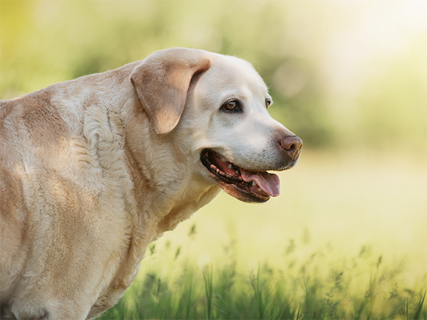
All Services
Learn More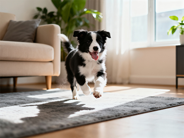
Keep
In Touch
© 2026 Treasure Coast Animal Rehabilitation & Fitness. All rights Reserved - Accessibility Statement - Privacy Policy - Sitemap
Powered by
